The Tonsils!
I decided to do my blog on the tonsils! I was trying to find a tissue that I normally take for granted and I came across the tonsils. The tonsils are lymphoid tissues that play a role in immune function which will be discussed further in this blog!
Role in Daily Life
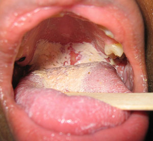
Usually, I don't think too much about my tonsils until I get sick and they become enlarged and inflamed (and cause me to be in pain). Or when I notice a white spots or mucus on them!

(Would definitely be making a trip to the doctor's office if this is what I saw!)
(Would definitely be making a trip to the doctor's office if this is what I saw!)
The tonsils play a role in the lymphatic system by detecting incoming pathogens in the mouth and nasal passages. They are secondary lymphoid structures and one of the Mucosa-Associated Lymphoid Tissues (other's include Peyer's patches and the Appendix)
The tissue of the tonsils contains B and T lymphocytes and some mature plasma cells but it's main role is mostly secretory function in the immune system and also helps to regulate immunoglobulin production.
The tonsils are most active under the age of ten and begin to involute after. The immune secretory function still remains as an adult but it occurs at lower levels than childhood (Mescher 2013).
Basic Anatomy
Did you know there are four types of tonsils? ( Most people only think there is one type of tonsil (the Palatine Tonsils that can be seen at the back of the throat)
They are Palatine, Lingual, Pharyngeal and Tubal Tonsils. Together, the tonsils compose of a structure called the Waldeyer's tonsillar ring (Roman 2011).
.jpg)
Figure A: Location of Pharyngeal, Tubal, Palatine and Lingual Tonsils which are located in the head and neck region (Together compose Waldeyer's Tonsillar ring)
Types of Tonsils
Palatine:
These are the largest of the four types and one palatine tonsil is located on each side of the soft palate. The palatine tonsil increases it's surface area through the use of tonsillar crypts which maximize the exposure to antigens and particles entering the body. These are deep invaginations lined with high amounts of lymphocytes and other types of leukocytes along the epithelial lining. Around the crypts are secondary lymphoid nodules. Each palatine tonsil has about 10 to 20 crypts (so 20- 40 between both) and has a thick partial capsule of dense connective tissue.Lingual:
These are small and numerous, located at the base of the tongue and have similar features to the Palatine tonsils. However each tonsil only has one crypt and no capsule forms over the tonsil.Pharyngeal:
There is only one pharyngeal tonsil and it is located in the nasopharynx passage. There are no tonsillar crypts but there are surface has shallow infoldings to increase the surface area. A thin partial connective tissue layer covers this tonsil (Paulsen 2010)Fun fact! When the pharyngeal tonsils are subject to chronic inflammation, regions become enlarged and are called the adenoid! ( Children with chronic ear infections often have these removed because enlargement can block the Eustachian tubes)

Tubal:
Two tubal tonsils surround the Eustachian tube openings in the ear (One on each tube). There is not as much information known about this type of tonsil.Histological Composition of the Tonsils
Palatine
Covered by non-keratinized stratified squamous epithelium. This type of tonsil is located at the back of the mouth, which is a moist cavity subject to abrasion due to the passage of food and other materials. This area is subject to abrasion so new cells line the basement membrane while older cells are pushed to the top.
Figure 1B: A section of the palatine tonsil, showing several secondary lymphoid nodules (LN), covered by a stratified squamous epithelium (E) and a capsule of connective tissue on the opposite side (CT). Germinal centers (GC) in the secondary lymphoid nodules can also be seen. Lumens of the crypts contain live and dead lymphocytes and bacteria. The stain used is H&E with a magnification of 140X

Figure 1C: Tonsillar crypts (C) are surrounded by stratified squamous epithelial cells (E) become infiltrated with lymphocytes and other leukocytes. Connective tissue in the top of the picture also contains many lymphocytes. A H&E stain is used with a magnification of 200X
Lingual
Covered by lightly keratinized stratified squamous epithelium. This type of tonsil is located at the base of the tongue, which is also an area of abrasion due to the passage of food. The keratin layer protects the mucosa from damage from mastication of food. The dorsal side tongue is also covered in a keratinized layer.Pharyngeal
Covered by ciliated pseudostratisfied columnar epithelium. The pharyngeal tonsil is located in the nasopharynx due to its location in the respiratory tract which is an area usually covered with this type of epithelium.Tubal
Covered by ciliated pseudostratisfied columnar epithelium. This is also located in the nasopharnyx area near the base of the Eustachian tubes (Mescher 2013).Pathologies
Normally the tonsils are colonized by aerobic and anaerobic species, which do not normally cause infections. However, these organisms and other pathogenic microbes can cause infections under the right circumstances. Some of these infections you have probably heard of (and if you're like me you've probably had one or more of these!)
Acute Pharyngotonsillitis
This can be caused by bacterial or viral (viral is more common) and patients symptoms include fever, malaise, painful swallowing and swollen lymph nodes.
Viral Infections:
Common viruses that cause this are adenovirus, rhinovirus, reovirus, respiratory syncytial virus (RSV) and influenza viruses. Epstein Barr Virus (a cause of infectious mononucleosis) also causes swollen and enlarged tonsils that are covered in a gray-white fluid.
Treatment includes analgesics like NSAIDs and acetaminophen, steroids in severe cases and lidocaine to relieve pain.

(You can see how swollen the left tonsil is!)
Bacterial Infections:
The most common cause of bacterial pharyngotonsillitis is cause by Group A β- hemolytic streptococcus. This is the group of bacteria that cause Strep throat- a common disease in children and teenagers. Symptoms of Strep throat include fever, sore throat, painful swallowing and swollen and enlarged tonsils and lymph nodes. When examining the tonsils, a whitish fluid (or spots) are also seen covering the tonsils (picture shown in the Role in Daily Life!)
Treatment includes antibiotics such as Penicillin V and Amoxicillin (and other macrolides for people allergic to Penicillin). Analgesics for pain relief are also commonly used such as NSAIDs and acetaminophen.
Fungal Infections:
In patients who are immunosuppressed or have undergone radiation treatment, Candida albicans (a opportunistic pathogen) can cause cottage cheese like plaques over pharyngeal mucosa.
Treatment includes oral nystatin preparations, lozenges and clotirimazole torches (Suurna 2012).
Recurrent Acute Tonsillitis
Some patients experience acute tonsillitis (multiple episodes of tonsillitis) with a complete recovery between episodes. Due to the tonsillar crypts, pathogens can easily become trapped in them causing recurrent infections.
Tonsillectomy (removal of the tonsils) is performed on patients with recurrent tonsillitis who have either 6-7 episodes in one year, 5 a year for two year or 3 a year for three years.

Chronic Tonsillitis
Patients with chronic tonsillitis are diagnosed when a sore throat occurs for more than three months with inflamed tonsils, bad breath and tenderness of lymph nodes.
Tonsilloliths are microbial biofilms which are the cause of bad breath and chronic tonsillitis in these patients. These appear as hard, white masses on the tonsils made up of debris such as live and dead microbes, food and other particles.
Tonsillectomy is also an treatment for chronic tonsillitis in adults (Mescher 2013).
References:
Information:Mescher A.L. (2013). Chapter 14. The Immune System & Lymphoid Organs. In A.L. Mescher (Ed), Junqueira’s Basic Histology, 13e. Retrieved October 23, 2013 from http://www.accessmedicine.com.qe2a-proxy.mun.ca/content.aspx?aID=57332245.
Paulsen D.F. (2010). Chapter 14. Lymphoid System. In D.F. Paulsen (Ed), Histology & Cell Biology: Examination & Board Review, 5e. Retrieved October 23, 2013 from http://www.accessmedicine.com.qe2a-proxy.mun.ca/content.aspx?aID=57094418.
Roman A.M. (2011). Chapter 28. Noninvasive Airway Management. In J.E. Tintinalli, J.S. Stapczynski, D.M. Cline, O.J. Ma, R.K. Cydulka, G.D. Meckler (Eds), Tintinalli's Emergency Medicine: A Comprehensive Study Guide, 7e. Retrieved October 23, 2013 from http://www.accessmedicine.com.qe2a-proxy.mun.ca/content.aspx?aID=6358328.
Suurna M.V. (2012). Chapter 21. Management of Adenotonsillar Disease. In A.K. Lalwani (Ed), CURRENT Diagnosis & Treatment in Otolaryngology—Head & Neck Surgery, 3e. Retrieved October 23, 2013 from http://www.accessmedicine.com.qe2a-proxy.mun.ca/content.aspx?aID=55767531.
Images:
http://web.uni-plovdiv.bg/stu1104541018/docs/res/skandalakis'%20surgical%20anatomy%20-%202004/Chapter%2013_%20Pharynx_fichiers/loadBinary(1).jpghttp://www.stdsandyou.com/yeastinfection/yeast-infection-of-the-mouth-candidiasis-oral-thrush.jpg
http://wacky5.com/wp-content/uploads/2010/05/unilateral-tonsillitis.jpg
http://www.edoctor.co.in/wp-content/uploads/2010/12/Tonsillitis.jpeg
https://blogger.googleusercontent.com/img/b/R29vZ2xl/AVvXsEiaLKbudpbt6bR-_2NKZXY9sxppOazRoWchHInMAD1_yQm6bE_RhHY_S6psVzlLZH23-3RrpG4ztHCEJOyaDRFPM9CbXoJqOmfU5vCIN9f-0HBwQ0obj90SDRDDmYoVKLkeso4uxLcpkIw/s1600/Tonsillectomy_tonsils.JPEG
I really appreciate DR AKHIGBE,my name is LAURIE HUGHES . I will never stop testifying DR AKHIGBE , Happiness is all i see now I never thought that I will be cured from HIV virus again. DR AKHIGBE did it for me I have been suffering from a deadly disease (HIV) for the past 2 years now, I had spent a lot of money going from one place to another, from churches to churches, hospitals have been my home every day residence. Constant checks up have been my hobby not until this faithful day, I saw a testimony on how DR AKHIGBE helped someone in curing his HIV disease in internet quickly I copied his email which is drrealakhigbe@gmail.com just to give him a test I spoke to him, he asked me to do some certain things which I did, he told me that he is going to provide the herbal cure to me, which he did, then he asked me to go for medical checkup after some days, after using the herbal cure and i did, behold I was free from the deadly disease,till now no HIV in me again he only asked me to post the testimony through the whole world, faithfully am doing it now,all the testimony of DR AKHIGBE is true please BROTHER and SISTER, MOTHER and FATHER he is great, I owe him in return. if you are having a similar problem just email him on drrealakhigbe@gmail.com or you can whats App his mobile number on +2348142454860 He can also cure these diseases like HIV and AIDS HERPES,DIABETICS,CANCER, HEPATITIS A&B,CHRONIC DISEASES, ASTHMA, HEART DISEASES, EXTERNAL INFECTION, EPILEPSY, STROKE, MULTIPLE SCLEROSIS, NAUSEA,VOMITING OR DIARRHEA,PARKINSON DISEASE,INFLUENZA,. COMMON COLD, AUTOIMMUNE DISORDER, LUPUS,ECZEMA,BACK PAIN, JOINT SCHIZOPHRENIA , PAIN.LOWER RESPIRATORY INFECTION. .ETC .please email drrealakhigbe@gmail.com or whats APP him ..+2348142454860 he is a real good and honest man.
ReplyDeletewebsite... https:drrealakhigbe.weebly.com
HERBAL CURE FOR SHINGLES,WARTS AND HERPES,HIV/AIDS
ReplyDelete5 years ago I had warts, I was treated with some liquid applied to the warts they continued to grow and spread... The next 2 doctors did laser surgery to remove them. 1 year after the surgery, they grew back close to where the 1st ones were' so I was finally told it was SHINGLES. I have had it for a very long time, I contracted it from my cheated boyfriend and I found out he was also infected and I ended the relationship between us. The warts were so embarrassed because it started spreading all over. I have been dealing with these things for a very long time. The last treatment I took was About 2 years ago I applied for natural treatment from Dr. JAMES HERBAL MIX MEDICINE, a week after I applied the treatment all the warts were gone. It's now 2 years and some months I don't have a single wart or any symptoms of SHINGLES. wow"" it's great, Dr. JAMES has finally cured me. If you know anyone suffering from these diseases or having any health challenges should contact Dr. JAMES for natural treatments. His herbal mix medicine is easy to drink with no side effects. Dr.James has cure for diseases like Pcos,Plantar-warts,Ovarian cyst, Parkinson's disease,Schizophrenia,Lung Cancer,Breast Cancer,Colo-Rectal Cancer,Blood Cancer,Prostate Cancer ,Epilepsy Dupuytren's disease ,Coeliac disease,Creutzfeldt–Jakob disease,Cerebral Amyloid Angiopathy, Ataxia,Arthritis,Amyotrophic Lateral Sclerosis,Fibromyalgia,Fluoroquinolone Toxicity
Syndrome Fibrodysplasia Ossificans ProgresS sclerosis,Seizures,Alzheimer's disease,Adrenocortical carcinoma.Asthma,Allergic diseases ,Copd,Glaucoma., Cataracts,Macular degeneration,Cardiovascular disease,Lung disease.Enlarged prostate,Osteoporosis,Lupus,Cushing’s disease,Heart failure,Multiple Sclerosis,Hypertension,Lyme Disease,Blood Cancer,Brain Cancer,Breast Cancer,Lung Cancer,Kidney Cancer, HIV/AIDS, Herpes Virus,Hepatitis B, Liver Inflammatory,Diabetes,Fibroid. Contact this Great man on his email and get a permanent cure from your diseases. Dr James Email Address [drjamesherbalmix@gmail.com].