Monday, November 11, 2013

Paper Review- Effect of adenoid hypertrophy on the voice and laryngeal mucosa in children

***This blog was designed as an assignment for the BIOL 3500 course at Memorial University of Newfoundland***


For the paper review, I chose the Gomaa et al. paper about the effect of hypertrophied (enlarged) adenoids on the voice and the mucosa of the larynx area in children.


Introduction:


In this study, Gomaa et al. look at the effects of hypertrophied adenoids in children, specifically the effects on voice and the laryngeal mucosa. As discussed in my previous blog, the adenoids (or pharyngeal tonsils) are located in the roof of the nasopharynx. When adenoids become infected, a 'drip' of the fluid can land on the mucosa of the vocal folds causing irritation (leading to coughing). Hypertrophied adenoids are common in children and cause symptoms such as snoring, nasal congestion, obstructive sleep apnea and recurrent ear infections. Dysphonia, defined an audible change in a person's voice (either detected by them or someone else), is a common occurrence in children ages 8 to 14. Dysphonia in children arises from voice abuse and misuse from chronic upper respiratory tract problems, such as hypertrophied adenoids, which can cause an impact on the voice-related quality of life.


Methods:


Patients were chosen from the Department of Otorhinolaryngology at the Faculty of Medicine, Minia University from Nov. 2011 to May 2012. Two groups of children were used- 40 children who suffered from adenoid hypertrophy without any other causes of upper respiratory disorders (the patient group) and 20 children in the control group who did not have any upper respiratory diseases or hypertrophied adenoids. 

Both groups of children had approximately the same mean age (6 to 7 years old) and the ratio of boys to girls used was about equal (slightly more girls in both groups). All subjects underwent a soft tissue X-rays as well as full history taking about history of dysphonia throughout life and family history of dysphonia or hearing loss.

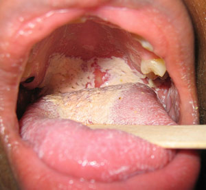
To assess patients, a laryngeal telescope and a test called the auditory perceptual assessment (APA) of voice was done using a modified GRBAS scale. The scale for dysphonia and voice quality used were the grade of dysphonia, strained voice, leaky voice, breathiness and irregularness. To evaluate the APA, researchers looked at the grade of dysphonia, character of the voice (if its strained, heavy, etc.), pitch and loudness.

Data collected was analyzed using SPSS version 17, with Chi square tests being performed between many proportions and a p-value of <0.05 was used for significance.


Results:


Dysphonia: 
Of the patient group, 6 had slight symptoms, 8 had moderate symptoms and the others did not show symptoms (35% of the patient group). The control group had only one child with slight symptoms. A p-value of 0.034 was determined showing an association between adenoid hypertrophy and dysphonia. 

Leaky Voice:
Twelve patients suffered from leaky voice (30%) while none of the control did, so a significant association was made between adenoid hypertrophy and leaky voice was apparent (p=0.024)

Pitch of Voice:
Decreased voice pitch was also associated with adenoid hypertrophy with a (p= 0.011) as 35% of the patient group suffered from this while only 5% of the control group did.

Laryngeal lesions:
Laryngeal lesions were found on 32.5% of the patient group and only 5% of the control group, so a statistical association was made between adenoid hypertrophy and laryngeal lesions

Strained voice, breathiness, irregularity in voice and the loudness of the voice did not show significance for an association with adenoid hypertrophy.

Table 2 from the publication showing the significance in the results obtained


https://mail-attachment.googleusercontent.com/attachment/u/1/?ui=2&ik=7034119b3e&view=att&th=14266de2d8efddac&attid=0.1&disp=inline&safe=1&zw&saduie=AG9B_P9iI_D0TshvYVSqE3pdONWi&sadet=1384705360983&sads=FAKNUPkhF0T2vJ9yrMD4_AlS4vA

Discussion:


The author discussed current research complementing the research performed in this study. Multiple studies show relationships between nasal problems/obstructions and dysphonia. However the results from this study disagree with a previous study by Simoes et al. which showed breathiness in deviant voices but not vocal strain in voices. In this study, neither a strained or breathy voices were found to be significant amongst patients with adenoid hypertrophy, so further research should be conducted to determine if there is significance.

The author also mentions that endoscopy is the gold standard for research such as this, but due to the cooperativeness of children for semi-invasive procedures, is harder to perform procedures like these.

A main point discussed is that hypertrophy of the adenoids can lead to a higher tension in the laryngeal muscle and increase the sub-glottal pressure during speech, which may be a reason for the occurrence of leaky voice, congestion in vocal folds, thickening of the mucosa and nodules in the vocal folds. This agrees with the results found in this study as dysphonia, leaky voice and laryngeal lesions were found to be significant in children with adenoid hypertrophy.

Another idea arises from turbulence in airflow from the enlargement of the adenoid causing a lower pressure which in turn causes the voice to be altered leading to dysphonia and a more nasal-sounding voice.

Overall the research performed showed the importance of assessment of voice and laryngeal examination in children with adenoid hypertrophy. It was also determined that laryngeal lesions should be treated once the adenoids are removed.


Personal Critique of Paper:


When reading this article, one of the first things I noticed was the small sample size. Sixty children were used in total with 40 in the patient group and 20 in the control group. While this isn't an overly small sample, to draw significance and to be applicable to other research this experiment should be repeated with more subjects.

There wasn't much histological discussion about the laryngeal mucosa or adenoid hypertrophy in this paper. In general, there doesn't appear to be much histological information and pictures about the adenoid in the literature I used. When discussing the damage to the laryngeal mucosa, this paper would have better exemplified that by showing a comparison between damaged and non damaged regions.

There was three tables presented in the paper, with only one of them showing results obtained. I think this paper could have been better explained with some more graphs and figures.

Another problem I encountered while reading the paper was the modified GRBAS scale used as I feel not enough clarification was given on what counted as slightly, moderate, severe, etc. The scale may also be subjective as these categories could be interpreted differently by different experimenters. I also felt that not enough information was given about the characteristics of voice. It is hard to determine what counts as leaky voice is for example as no background is given for the four characteristics of voice. Overall, I found that the terms were not well defined, leading me to to more research about the topic.

I also thought there should have been more information on laryngeal lesions as they mentioned how there was a statistical association between them and adenoid hypertrophy. They mentioned minimal lesions are caused by adenoid hypertrophy and the importance of treating them, but this doesn't give the reader a clear idea on the reasoning of their importance. Also, the wording appears to be incorrect as on one page the author talks about how 32.5% of patients have laryngeal lesions and only 5% of the control does, while at the end, they say that laryngeal lesions only happen with adenoid hypertrophy, which doesn't support the previous statement.

Overall, I felt this paper needed some more clarification on how the scales were determined to test the subjects as well as clearer wording as I felt there was confusion with some main points.


Tuesday, October 22, 2013

Histology of the Tonsils (And a bit of Everything Else!)

This blog was designed as an assignment for the BIOL 3500 course at Memorial University of Newfoundland


The Tonsils!

I decided to do my blog on the tonsils! I was trying to find a tissue that I normally take for granted and I came across the tonsils. The tonsils are lymphoid tissues that play a role in immune function which will be discussed further in this blog!


Role in Daily Life

Usually, I don't think too much about my tonsils until I get sick and they become enlarged and inflamed (and cause me to be in pain). Or when I notice a white spots or mucus on them!


A set of large tonsils in the back of the throat covered in white exudate


(Would definitely be making a trip to the doctor's office if this is what I saw!)

The tonsils play a role in the lymphatic system by detecting incoming pathogens in the mouth and nasal passages. They are secondary lymphoid structures and one of the Mucosa-Associated Lymphoid Tissues (other's include Peyer's patches and the Appendix)

The tissue of the tonsils contains B and T lymphocytes and some mature plasma cells but it's main role is mostly secretory function in the immune system and also helps to regulate immunoglobulin production.

The tonsils are most active under the age of ten and begin to involute after. The immune secretory function still remains as an adult but it occurs at lower levels than childhood (Mescher 2013).


Basic Anatomy

Did you know there are four types of tonsils? ( Most people only think there is one type of tonsil (the Palatine Tonsils that can be seen at the back of the throat)

They are Palatine, Lingual, Pharyngeal and Tubal Tonsils. Together, the tonsils compose of a structure called the Waldeyer's tonsillar ring (Roman 2011).


Figure A: Location of Pharyngeal, Tubal, Palatine and Lingual Tonsils which are located in the head and neck region (Together compose Waldeyer's Tonsillar ring)


Types of Tonsils


Palatine:

These are the largest of the four types and one palatine tonsil is located on each side of the soft palate. The palatine tonsil increases it's surface area through the use of tonsillar crypts which maximize the exposure to antigens and particles entering the body. These are deep invaginations lined with high amounts of lymphocytes and other types of leukocytes along the epithelial lining. Around the crypts are secondary lymphoid nodules. Each palatine tonsil has about 10 to 20 crypts (so 20- 40 between both) and has a thick partial capsule of dense connective tissue.

Lingual: 

These are small and numerous, located at the base of the tongue and have similar features to the Palatine tonsils. However each tonsil only has one crypt and no capsule forms over the tonsil.


Pharyngeal: 

There is only one pharyngeal tonsil and it is located in the nasopharynx passage. There are no tonsillar crypts but there are surface has shallow infoldings to increase the surface area. A thin partial connective tissue layer covers this tonsil (Paulsen 2010)

Fun fact!   When the pharyngeal tonsils are subject to chronic inflammation, regions become enlarged and are called the adenoid! ( Children with chronic ear infections often have these removed because enlargement can block the Eustachian tubes)


Tubal:

Two tubal tonsils surround the Eustachian tube openings in the ear (One on each tube). There is not as much information known about this type of tonsil.


Histological Composition of the Tonsils


Palatine  

Covered by non-keratinized stratified squamous epithelium. This type of tonsil is located at the back of the mouth, which is a moist cavity subject to abrasion due to the passage of food and other materials. This area is subject to abrasion so new cells line the basement membrane while older cells are pushed to the top.


Figure 1B: A section of the palatine tonsil, showing several secondary lymphoid nodules (LN), covered by a stratified squamous epithelium (E) and a capsule of connective tissue on the opposite side (CT). Germinal centers (GC) in the secondary lymphoid nodules can also be seen. Lumens of the crypts contain live and dead lymphocytes and bacteria. The stain used is H&E with a magnification of 140X


Figure 1C: Tonsillar crypts (C) are surrounded by stratified squamous epithelial cells (E) become infiltrated with lymphocytes and other leukocytes. Connective tissue in the top of the picture also contains many lymphocytes. A H&E stain is used with a magnification of 200X


Lingual  

Covered by lightly keratinized stratified squamous epithelium. This type of tonsil is located at the base of the tongue, which is also an area of abrasion due to the passage of food. The keratin layer protects the mucosa from damage from mastication of food. The dorsal side tongue is also covered in a keratinized layer.


Pharyngeal

Covered by ciliated pseudostratisfied columnar epithelium. The pharyngeal tonsil is located in the nasopharynx due to its location in the respiratory tract which is an area usually covered with this type of epithelium.


Tubal

Covered by ciliated pseudostratisfied columnar epithelium. This is also located in the nasopharnyx area near the base of the Eustachian tubes (Mescher 2013).


Pathologies


Normally the tonsils are colonized by aerobic and anaerobic species, which do not normally cause infections. However, these organisms and other pathogenic microbes can cause infections under the right circumstances. Some of these infections you have probably heard of (and if you're like me you've probably had one or more of these!)


Acute Pharyngotonsillitis


This can be caused by bacterial or viral (viral is more common) and patients symptoms include fever, malaise, painful swallowing and swollen lymph nodes. 


Viral Infections:

Common viruses that cause this are adenovirus, rhinovirus, reovirus, respiratory syncytial virus (RSV) and influenza viruses. Epstein Barr Virus (a cause of infectious mononucleosis) also causes swollen and enlarged tonsils that are covered in a gray-white fluid.
Treatment includes analgesics like NSAIDs and acetaminophen, steroids in severe cases and lidocaine to relieve pain.


(You can see how swollen the left tonsil is!)

Bacterial Infections:

The most common cause of bacterial pharyngotonsillitis is cause by Group A β- hemolytic streptococcus. This is the group of bacteria that cause Strep throat- a common disease in children and teenagers. Symptoms of Strep throat include fever, sore throat, painful swallowing and swollen and enlarged tonsils and lymph nodes. When examining the tonsils, a whitish fluid (or spots) are also seen covering the tonsils (picture shown in the Role in Daily Life!)
Treatment includes antibiotics such as Penicillin V  and Amoxicillin (and other macrolides for people allergic to Penicillin). Analgesics for pain relief are also commonly used such as NSAIDs and acetaminophen.


Fungal Infections:

In patients who are immunosuppressed or have undergone radiation treatment, Candida albicans (a opportunistic pathogen) can cause cottage cheese like plaques over pharyngeal mucosa.

Treatment includes oral nystatin preparations, lozenges and clotirimazole torches
(Suurna 2012).


Recurrent Acute Tonsillitis


Some patients experience acute tonsillitis (multiple episodes of tonsillitis) with a complete recovery between episodes. Due to the tonsillar crypts, pathogens can easily become trapped in them causing recurrent infections.


Tonsillectomy (removal of the tonsils) is performed on patients with recurrent tonsillitis who have either 6-7 episodes in one year, 5 a year for two year or 3 a year for three years. 


Chronic Tonsillitis


Patients with chronic tonsillitis are diagnosed when a sore throat occurs for more than three months with inflamed tonsils, bad breath and tenderness of lymph nodes.

Tonsilloliths are microbial biofilms which are the cause of bad breath and chronic tonsillitis in these patients. These appear as hard, white masses on the tonsils made up of debris such as live and dead microbes, food and other particles.

Tonsillectomy is also an treatment for chronic tonsillitis in adults
(Mescher 2013).

References:

Information:

Mescher A.L. (2013). Chapter 14. The Immune System & Lymphoid Organs. In A.L. Mescher (Ed), Junqueira’s Basic Histology, 13e. Retrieved October 23, 2013 from http://www.accessmedicine.com.qe2a-proxy.mun.ca/content.aspx?aID=57332245.

Paulsen D.F. (2010). Chapter 14. Lymphoid System. In D.F. Paulsen (Ed), Histology & Cell Biology: Examination & Board Review, 5e. Retrieved October 23, 2013 from http://www.accessmedicine.com.qe2a-proxy.mun.ca/content.aspx?aID=57094418.

Roman A.M. (2011). Chapter 28. Noninvasive Airway Management. In J.E. Tintinalli, J.S. Stapczynski, D.M. Cline, O.J. Ma, R.K. Cydulka, G.D. Meckler (Eds), Tintinalli's Emergency Medicine: A Comprehensive Study Guide, 7e. Retrieved October 23, 2013 from http://www.accessmedicine.com.qe2a-proxy.mun.ca/content.aspx?aID=6358328.


Suurna M.V. (2012). Chapter 21. Management of Adenotonsillar Disease. In A.K. Lalwani (Ed), CURRENT Diagnosis & Treatment in Otolaryngology—Head & Neck Surgery, 3e. Retrieved October 23, 2013 from http://www.accessmedicine.com.qe2a-proxy.mun.ca/content.aspx?aID=55767531.


Images:

http://web.uni-plovdiv.bg/stu1104541018/docs/res/skandalakis'%20surgical%20anatomy%20-%202004/Chapter%2013_%20Pharynx_fichiers/loadBinary(1).jpg

http://www.stdsandyou.com/yeastinfection/yeast-infection-of-the-mouth-candidiasis-oral-thrush.jpg

http://wacky5.com/wp-content/uploads/2010/05/unilateral-tonsillitis.jpg

http://www.edoctor.co.in/wp-content/uploads/2010/12/Tonsillitis.jpeg

https://blogger.googleusercontent.com/img/b/R29vZ2xl/AVvXsEiaLKbudpbt6bR-_2NKZXY9sxppOazRoWchHInMAD1_yQm6bE_RhHY_S6psVzlLZH23-3RrpG4ztHCEJOyaDRFPM9CbXoJqOmfU5vCIN9f-0HBwQ0obj90SDRDDmYoVKLkeso4uxLcpkIw/s1600/Tonsillectomy_tonsils.JPEG